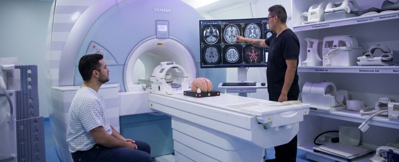
Brain MRI Scan: What It Detects, How It Works & What to Expect
What Is a Brain MRI Scan?
MRI stands for Magnetic Resonance Imaging. MRI scans do not involve X-ray technology, nor do they involve ionizing radiation. Instead, MRI scans rely upon strong magnets and radio waves to develop very high-resolution images of the internal structures of the body.
When it comes to diagnosing and monitoring many forms of neurological conditions (including brain tumors and strokes), MRI has become the gold-standard imaging tool. This is because MRI can reveal abnormalities in both the brain's structural integrity and its functional processes with much greater detail than either X-rays or CT scans.
What Does a Brain MRI Detect?
An MRI of the brain can detect a variety of abnormalities in the brain. These include hemorrhages, abscesses, cysts, arteriovenous malformations, and other lesions. An MRI can also reveal damage caused by traumatic brain injuries. In addition, an MRI can show evidence of certain diseases affecting the brain and central nervous system. Examples of these diseases include multiple sclerosis, Parkinson's disease, Alzheimer's disease, Huntington's disease, amyotrophic lateral sclerosis (ALS), and many others.
Types of Brain MRI Scans
There are two primary types of brain MRI scans:
Standard Brain MRI:
This type of MRI uses magnetic fields and radio waves alone to develop images of the brain and the surrounding structures. Most patients who undergo a brain MRI will receive this type of scan, which does not require any injected contrast material.
Brain MRI With Contrast:
Contrast-enhanced brain MRI is performed using an intravenous injection of a contrast agent, most commonly gadolinium-based, administered just before or during the actual scan. By altering the magnetic properties of nearby water molecules, gadolinium enhances image clarity and allows for better detection of various pathological states.
The decision of whether to perform an MRI with contrast depends on the patient's specific clinical situation, such as the following:
- Is there a tumor?
- Is it likely to contain blood vessels?
- Will the presence of inflammation affect the appearance of a tumor?
- Does the patient have diabetes mellitus or kidney dysfunction?
- Is there a history of allergic reactions to iodine-containing substances?
- Has the patient received previous injections of gadolinium-based agents?
Also read: Brain tumours type
How to Prepare for a Brain MRI
Preparation for a brain MRI is relatively simple and easy. Typically, you can eat and drink as usual before undergoing a brain MRI unless instructed differently by your physician. Also, you can continue taking your prescribed medications unless instructed differently by your physician.
However, there are several important things to keep in mind when preparing for a brain MRI:
- Are you claustrophobic?
- Do you have trouble lying flat or staying motionless for extended periods?
- Are you having difficulty swallowing due to recent surgery?
- Do you have metal fragments lodged in your head or eye(s)?
- Do you have a pacemaker?
- Do you have a vagus nerve stimulator?
- Do you have any other type of implantable medical device?
It is also a good idea to let your healthcare provider know ahead of time if you are pregnant. Wear loose-fitting clothing with no metal fasteners. Remove all jewelry and metal items before entering the MRI suite, including watches, earrings, glasses frames, etc. Remove all hearing aids, hairpins, underwire bras, dentures, dental implants, and prosthetic limbs. Remove all electronic devices, including cell phones.
What Happens During a Brain MRI Scan?
The actual process of getting a brain MRI is painless and easy. When you arrive for your appointment, you will be asked to change into a hospital gown and lie down on the MRI scanning table. The technician will place a "head coil" helmet around your head to improve image clarity.
Some head coils contain mirrors or small screens that allow you to watch movies while being scanned to minimize anxiety related to confined spaces.
As soon as you are positioned properly within the MRI machine, the technician will begin capturing images of your brain. You will hear loud knocking and clicking noises from the machine during the scanning process. However, you will wear ear protection to prevent damage to your eardrums.
You may also choose to listen to music during the scanning process. Since movement during the scanning process could degrade image quality, please try to remain as still as possible.
Since you will be able to communicate verbally with the technician at all times via an intercom system, please do not hesitate to ask for assistance if needed. Additionally, please hold onto the call button provided so that we can quickly assist you in case of an emergency.
What to Expect After a Brain MRI
Typically, after completing the brain MRI, you can resume your daily routine immediately, unless you received sedation before the procedure; in that case, you should arrange for someone to escort you home.
Generally speaking, there is no recovery time for a standard brain MRI. After receiving contrast during your brain MRI, you should drink lots of fluids to help flush out the contrast agent from your body. Minor bruising from the IV site should disappear within three days.
Understanding Brain MRI Results
After completing your brain MRI, our radiologists review your images and write a report detailing their findings. They then send a copy of your report to your referring physician within one to two business days. Based on their interpretation of your images and findings in conjunction with your symptoms and medical history, your physician will then schedule a consultation with you to go over what the findings indicate regarding your potential diagnosis or future course of treatment.
Also read: Brain tumour treatment in UAE
Brain MRI vs. Other Imaging Tests
Magnetic resonance imaging (MRI) is generally regarded as the most sensitive form of imaging available today to evaluate the structure and function of the brain. While CT scans are faster and more widely available than MRI scanners (making them more attractive options for emergency procedures such as evaluating suspected stroke or head trauma), they do not provide anywhere near as detailed an evaluation of soft tissue in the same way that MRI does.
Brain MRI Scan FAQs
How many hours does a brain MRI take?
Can MRI results be seen immediately?
What to avoid during a brain MRI?
Why would a neurologist order a brain MRI?
Does a brain MRI detect all brain conditions?
Can I eat before a brain MRI?
Can a brain MRI detect a stroke?
References:
- Braffman, B. H., Zimmerman, R. A., Trojanowski, J. Q., Gonatas, N. K., Hickey, W. F., & Schlaepfer, W. W. (1988). Brain MR: Pathologic correlation with gross and histopathology. American Journal of Roentgenology, 151(3), 551–558.
- Filippi, M., Agosta, F., Barkhof, F., Dubois, B., Fox, N. C., Frisoni, G. B., Jack, C. R., Johannsen, P., Miller, B. L., Nestor, P. J., Scheltens, P., Sorbi, S., Teipel, S., Thompson, P. M., & Wahlund, L. O. (2012). EFNS task force: The use of neuroimaging in the diagnosis of dementia. European Journal of Neurology, 19(12), e131–e140.
- Gaillard, W. D., Chiron, C., Cross, J. H., Harvey, A. S., Kuzniecky, R., Hertz-Pannier, L., & Vezina, L. G. (2009). Guidelines for imaging infants and children with recent-onset epilepsy. Epilepsia, 50(9), 2147–2153.
- Schellinger, P. D., Bryan, R. N., Caplan, L. R., Detre, J. A., Edelman, R. R., Jaigobin, C., Kidwell, C. S., Mohr, J. P., Sloan, M., Sorensen, A. G., & Warach, S. (2010). Evidence-based guideline: The role of diffusion and perfusion MRI for the diagnosis of acute ischemic stroke. Neurology, 75(2), 177–185.
- Tsao, J. (2010). Ultrafast imaging: Principles, pitfalls, solutions, and applications. Journal of Magnetic Resonance Imaging, 32(2), 252–266.
Need to consult a specialist?